Download the 7-step guide:
The new 3.1 All-In-One TBS Report is divided into 7 steps. In the next few days, we will show you how easy it is to use the new report in daily clinical routine.
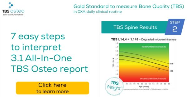
Today – STEP 2 of 7 – TBS spine results
Check the bone microarchitecture of the patient. The Trabecular Bone Score (TBS) result is plotted onto a reference graph. The Trabecular Bone Score (TBS) normal values according to age are represented by the thick black line. The thinner lines above and below represent this normative curve +/- 1 SD (standard deviation).
A gradient of different-color zones representing different status of bone microarchitecture: high TBS values (TBS L1-L4 > 1.31) representing Normal microarchitecture, and low TBS values (TBS L1-L4 ≤ 1.23) representing Degraded microarchitecture.
➢ Why is this important?
- With this graph, you can see how the TBS score of the patient compares to the normal population (same age, same gender, same ethnicity) and see if the patient is at high risk of fracture based on the microarchitecture assessment only.
➢ What to do?
- Use the colors to assess your patient’s fracture risk based on TBS:
- Green zone: low risk of fracture, suggesting normal bone microarchitecture.
- Yellow zone: medium risk of fracture, suggesting partially degraded bone
microarchitecture. - Red zone: high risk of fracture, suggesting degraded bone microarchitecture.
Feel free to share with your colleagues, as it helps identify more patients at risk for fracture. If you
have questions, contact us at support@medimapsgroup.com – we will gladly help you.
Download the 7-step guide here:
As a reminder the main features of the 3.1 All-In-One TBS Osteo report are:
- Completely new and intuitive design to simplify the interpretation of the BMD and TBS exam.
- Bone health report – All-In-One: TBS and BMD results in one report
- New BMD T-score adjusted for TBS
- Fracture risk grid – A visualization of BMD and TBS combined assessment
- Automatic and editable conclusion developed by international experts to communicate easily with referring doctors
Feel free to share with your colleagues, as it helps identify more patients at risk for fracture. If you have questions, contact us at support@medimapsgroup.com – we will gladly help you.
Click here to follow us on LinkedIn.
You can find more information on all 7 steps below: